


Diabetic retinopathy, the most common diabetic eye disease, occurs when there are changes in the blood vessels in the retina. Sometimes, these vessels can swell and let out fluids, or even cover completely. In other cases, new abnormal blood vessels grow on the surface of the retina.
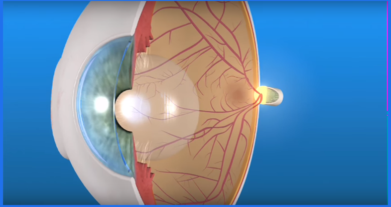
The retina is a thin layer of light-sensitive tissue that covers the back of the eye. The light rays focus on the retina, where they are transmitted to the brain and interpreted as images. The macula is a very small area in the center of the retina. The macula is the area responsible for detailed vision, allowing us to read, sew or recognize a face. The part around the retina, called the peripheral retina, is responsible for lateral or peripheral vision.
Generally, diabetic retinopathy affects both eyes. People with diabetic retinopathy often do not realize changes in their vision during the early stages of the disease. But as it progresses, diabetic retinopathy usually causes a loss of vision that in many cases cannot be reversed.
Diabetic eye problems
There are two types of diabetic retinopathy:
There are two types of diabetic retinopathy:
Nonproliferative diabetic retinopathy (RDNP) is the earliest stage of diabetic retinopathy. When this condition exists, impaired blood vessels allow an escape of blood fluids inside the eye. Occasionally, deposits of cholesterol or other blood fats may enter the retina.
RDNP can cause changes in the eyes, including:
Microaneurysms: Small bumps in the blood vessels of the retina that often leak fluid.
Hemorrhages of the retina: Small blood spots that enter the retina.
Hard exudatest is the inflammation or thickening of the macula due to fluid leaks from the blood vessels of the retina. The macula does not work properly when it is inflamed. Macular edema is the most common cause of vision loss during diabetes.
Macular exudates: t is the inflammation or thickening of the macula due to fluid leaks from the blood vessels of the retina. The macula does not work properly when it is inflamed. Macular edema is the most common cause of vision loss during diabetes.
Macular ischemiaSmall blood vessels (capillaries) close or plug. Your vision becomes blurred because the macula does not receive enough blood to function properly.
Many people with diabetes have mild RDNP, which usually does not affect vision. However, if your vision is affected, it is as a result of macular edema and macular ischemia.
Notice how macular edema and macular ischemia affect the eyes
Proliferative Diabetic Retinopathy (RDP)
Proliferative diabetic retinopathy (RDP) occurs mainly when many of the blood vessels in the retina become clogged, preventing sufficient blood flow. In an attempt to supply blood to the area where the original vessels have become clogged, the retina responds by creating new blood vessels. This process is called neovascularization. However, the new blood vessels are also abnormal and do not provide the retina with adequate blood flow. Often, the new vessels are accompanied by scar tissue that can cause the retina to wrinkle or detach.
RDP can cause more severe vision loss than RDNP, as it can affect both central and peripheral vision. RDP affects vision in the following ways:
Vitreous hemorrhage: The new and delicate blood vessels bleed into the vitreous (the gelatinous substance in the center of the eye), preventing light rays from reaching the retina. If the bleeding is small, you may see some new dark and floating spots. A very large hemorrhage can block vision, allowing you to only see the difference between light and dark. Vitreous hemorrhage alone does not cause permanent vision loss. Whenever the blood disappears, vision can return to its previous state, unless the macula has been damaged.
Traction retinal detachment: When the tissue of a scar caused by neovascularization shrinks, the retina wrinkles and can detach from its normal position. These macular wrinkles can distort vision. More severe vision loss may occur if the macula or large areas of the retina detach.
Neovascular glaucoma: If a series of retinal vessels close, a neovascularization in the iris (the colored part of the eye) may occur. When this condition exists, new blood vessels can block the normal flow of fluid in the eye. The pressure in the eye increases, which presents a particularly serious condition that causes damage to the optic nerve.
Notice how a vitreous hemorrhage affects your eyes.
Article written by the American Academy of Ophthalmology